Native peptides possess high target affinity and low toxicity, making them valuable tools for biochemical assays and structural biology. However, their utility in prolonged laboratory experiments or in vivo models is frequently restricted by poor enzymatic stability, rapid renal clearance, and structural flexibility. Peptide modification directly addresses these inherent limitations. By systematically altering the chemical structure of a specific sequence, researchers can optimize pharmacokinetic properties, enforce conformational rigidity, and attach functional probes for specific analytical assays. This guide examines standard and advanced peptide modifications, their biochemical mechanisms, and their precise applications in laboratory research.
The Role of Modifications in Peptide Engineering
Unmodified linear sequences are highly susceptible to proteolytic degradation in serum or intracellular environments. Exopeptidases cleave amino acids from the exposed termini, while endopeptidases hydrolyze internal peptide bonds. Furthermore, short linear peptides often exist as an ensemble of conformations in solution. This lack of structural constraint reduces the thermodynamic favorability of receptor binding, as significant entropy is lost upon adopting the active conformation. Structural and terminal modifications are engineered to resolve these issues, converting transient sequences into robust experimental probes.
N-Terminal and C-Terminal Modifications
The most immediate vulnerabilities on any linear peptide are its free N-terminal amine and C-terminal carboxylic acid. Shielding these ends is the fundamental first step in increasing sequence half-life.
Acetylation (N-terminus)
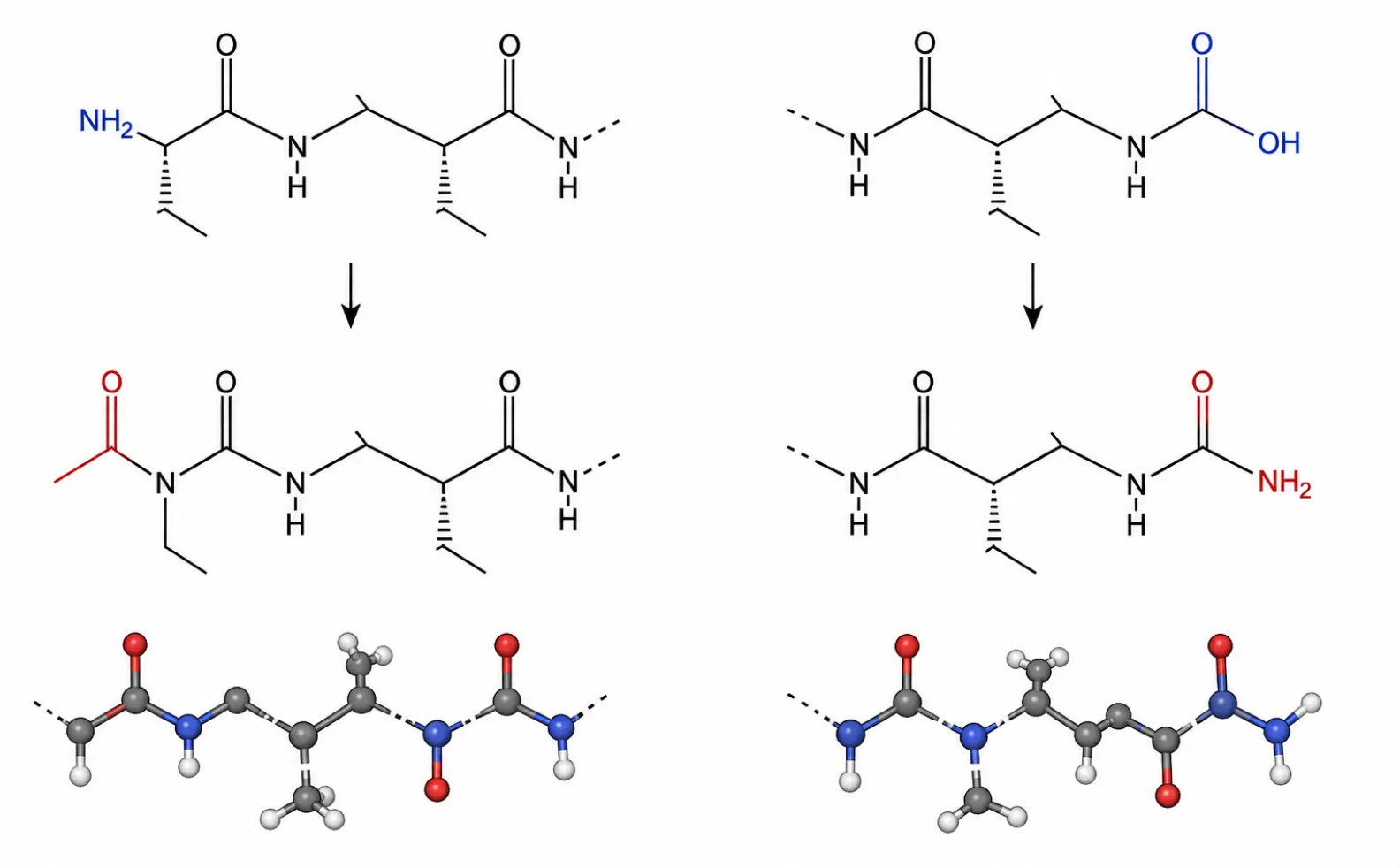
Acetylation involves the attachment of an acetyl group to the N-terminal amine, removing its positive charge and replacing it with a neutral amide bond. This modification mimics the natural state of many intracellular proteins and completely blocks the action of N-terminal aminopeptidases. It is a standard requirement for peptides intended for cellular assays where serum exposure is unavoidable.
Amidation (C-terminus)
Amidation replaces the negatively charged C-terminal hydroxyl group with a neutral amide group. This prevents recognition by carboxypeptidases. When a peptide is both N-acetylated and C-amidated, it is protected from terminal degradation. Additionally, neutralizing both termini removes unnatural charges from the ends of the sequence, allowing the peptide to fold or bind without electrostatic interference from its own backbone boundaries.
Structural Modifications for Stability and Affinity
To prevent internal enzymatic cleavage and lock a peptide into a specific bioactive shape, researchers utilize structural constraints. The most prominent methodology is cyclization.
Peptide Cyclization
Cyclization restricts the conformational freedom of a sequence. This physical constraint shields internal bonds from endopeptidases and pre-organizes the peptide into its binding conformation, which typically increases receptor affinity by several orders of magnitude. For specific constrained sequence synthesis, researchers rely on cyclopeptides.
- Disulfide Bridges: The most common form of cyclization involves the oxidation of the thiol groups on two cysteine residues to form a covalent disulfide bond. While straightforward to synthesize, disulfide bonds are susceptible to reduction in highly reducing intracellular environments.
- Amide (Lactam) Bridges: Formed by condensing the side chains of specific amino acids (typically the amine of Lysine and the carboxylic acid of Glutamic Acid or Aspartic Acid), lactam bridges create a chemically stable ring structure that resists reduction, offering superior stability for in vivo models.
- Head-to-Tail Cyclization: This method links the N-terminus directly to the C-terminus, creating a continuous loop. It completely eliminates terminal exopeptidase targets and provides maximum conformational rigidity.
PEGylation
Conjugation with Polyethylene Glycol (PEG) is utilized to increase the molecular weight and hydrodynamic radius of a peptide. The bulky, hydrated PEG polymer creates a steric shield around the peptide backbone, physically blocking proteases from accessing cleavage sites. Additionally, increasing the mass of the conjugate above the renal filtration threshold (~40-50 kDa) drastically reduces clearance rates. PEGylation requires precise site selection; attaching the polymer too close to the active binding domain will result in steric hindrance and a loss of biological activity.
Functional and Analytical Modifications
Beyond stability, modifications are used to attach functional tags for tracking, purification, and interaction mapping.
Fluorescent Labeling
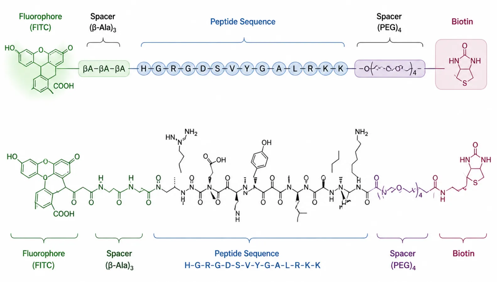
For applications in flow cytometry, fluorescence microscopy, and binding kinetics, fluorophores are covalently conjugated to the peptide. FITC (Fluorescein isothiocyanate) and FAM are standard choices for green emission, while TAMRA and Cyanine dyes (Cy3, Cy5) provide red to near-infrared options. To prevent the bulky hydrophobic fluorophore from interfering with the peptide’s native binding behavior, it is standard practice to insert an aliphatic spacer, such as Ahx (aminohexanoic acid) or a short PEG chain, between the peptide sequence and the dye.
Biotinylation
The non-covalent interaction between biotin and streptavidin (or avidin) is one of the strongest known in nature, with a dissociation constant (Kd) on the order of 10^-14 mol/L. Biotinylated peptides are critical for pull-down assays, surface plasmon resonance (SPR) immobilization, and enzyme-linked immunosorbent assays (ELISA). Similar to fluorescent tags, biotin is typically attached via an Ahx or PEG spacer to ensure the tag remains accessible to the bulky streptavidin tetramer once the peptide is bound to its target.
Phosphorylation
Signal transduction pathways are largely regulated by the reversible phosphorylation of serine, threonine, and tyrosine residues. The synthesis of specifically phosphorylated peptides enables researchers to map kinase specificity, determine phosphatase kinetics, and isolate phospho-dependent binding proteins (such as those containing SH2 domains). Incorporating phosphorylated residues requires specialized building blocks during synthesis and careful handling to prevent dephosphorylation during cleavage and purification.
Synthesis and Manufacturing Complexities
Introducing complex modifications during Solid-Phase Peptide Synthesis (SPPS) requires rigorous chemical strategies, specifically orthogonal protection. In standard SPPS, side chains are protected by groups that are stable to the iterative Fmoc deprotection cycles but are cleaved simultaneously with the final detachment of the peptide from the resin.
When a modification (such as a lactam bridge or a specific side-chain fluorophore) is required, the target residues must be protected by distinct, "orthogonal" groups that can be selectively removed while the rest of the sequence remains protected and anchored to the resin. For example, synthesizing a side-chain to side-chain lactam bridge often utilizes Allyl and Alloc protecting groups, which are selectively removed using a palladium catalyst before the final cleavage step. These additional on-resin reactions increase the complexity of the synthesis, decrease the overall yield, and demand high-resolution analytical validation via HPLC and mass spectrometry to confirm absolute correct mass and purity. Partnering with a specialized facility for peptide modification services ensures these intricate chemical requirements are met without compromising the integrity of the final product.
Selecting the Optimal Modification Strategy
The choice of modification must strictly align with the biological objective of the experiment. For simple in vitro enzymatic assays, terminal acetylation and amidation may provide sufficient stability. For robust animal models or complex pharmacokinetic studies, cyclization or PEGylation is often mandatory. When designing probes for imaging or pull-down assays, the placement of the fluorophore or biotin tag—and the inclusion of an appropriate spacer—determines the functional viability of the construct. For sequences that demand multiple simultaneous modifications or non-standard conjugations, our custom peptide synthesis platform provides the analytical precision required for advanced research applications.
FAQ
What is the functional difference between N-terminal acetylation and C-terminal amidation?
Acetylation neutralizes the positive charge of the N-terminus, while amidation neutralizes the negative charge of the C-terminus. Functionally, acetylation blocks N-terminal aminopeptidases, and amidation blocks C-terminal carboxypeptidases. Using both modifications together provides complete protection against terminal enzymatic degradation in biological sera.
How does cyclization improve the binding affinity of a peptide?
Linear peptides lose significant entropy when they fold into a bioactive conformation upon binding a receptor. Cyclization pre-organizes the peptide into this rigid conformation before binding occurs. Because less thermodynamic energy is spent restricting the molecule’s shape during the binding event, the overall binding affinity is typically increased.
Why is a spacer necessary when attaching a fluorescent tag or biotin?
Fluorophores and biotin are bulky molecules. If attached directly to the peptide sequence, they can cause steric hindrance, physically blocking the peptide from interacting with its target receptor. A flexible spacer, such as Ahx (aminohexanoic acid) or a PEG chain, provides distance between the functional tag and the active peptide domain, preserving biological activity.
Does PEGylation negatively impact peptide activity?
It can, if improperly designed. While PEGylation significantly increases half-life and prevents proteolytic degradation by shielding the peptide, the large polymer chain can also block receptor binding. Site-specific PEGylation, placed away from the known binding interface, is required to balance improved pharmacokinetics with retained biological function.
Why are orthogonal protecting groups required for peptide cyclization?
Orthogonal protecting groups can be removed under specific chemical conditions that do not affect the main protecting groups on the rest of the peptide. This allows chemists to selectively expose only the two side chains intended for cyclization (e.g., forming a lactam bridge) while the rest of the sequence remains protected, preventing unwanted cross-reactions during synthesis.
Can multiple modifications be applied to a single synthetic peptide?
Yes, multiple modifications are routinely combined. For example, a single sequence can be N-acetylated, C-amidated, internally cyclized via a disulfide bond, and conjugated with a fluorophore on a specific lysine residue. However, each additional modification increases synthesis complexity, requires more sophisticated orthogonal protection strategies, and generally reduces the final overall yield.
-

Synpeptide
peptide-focused CRO/CDMO company
The SynPeptide Research Team brings together scientists specializing in peptide synthesis, purification, and analytical characterization. Drawing on hands-on laboratory experience across custom and catalog peptides, the team shares evidence-based insights for researchers, formulators, and product developers. All content is reviewed against current scientific literature and internal quality-control data, reflecting SynPeptide's commitment to accuracy, reproducibility, and the responsible communication of peptide science.