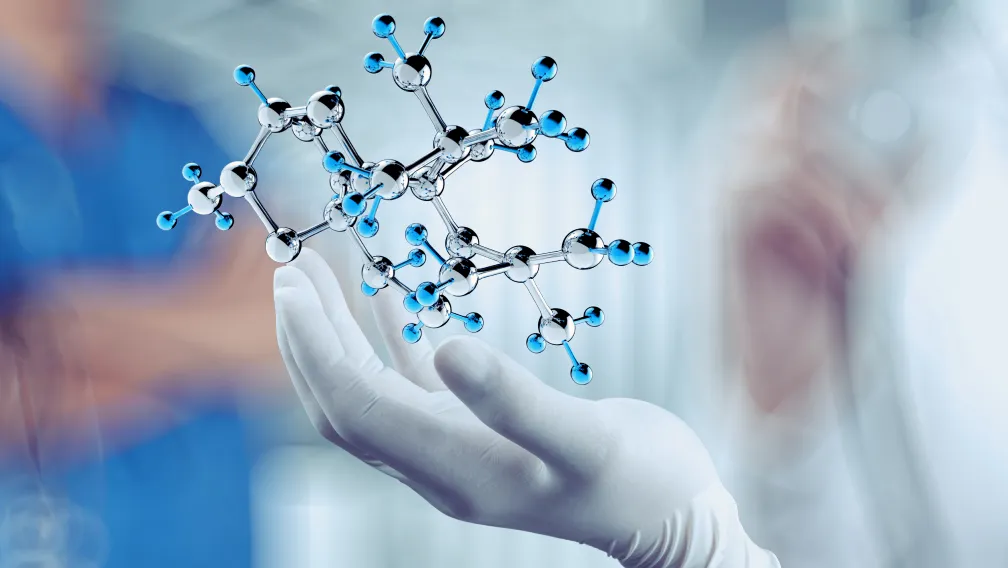
In the field of molecular biology and synthetic chemistry, peptide nucleic acids (PNAs) represent a critical advancement in oligonucleotide engineering. Originally conceptualized in the early 1990s, PNAs are synthetic DNA/RNA analogs characterized by an uncharged, achiral, pseudopeptide backbone. Unlike traditional oligonucleotides, which rely on a negatively charged sugar-phosphate backbone, PNAs utilize repeating N-(2-aminoethyl)glycine units linked by peptide bonds. The nucleobases (adenine, cytosine, guanine, and thymine) are tethered to this backbone via methylenecarbonyl linkages. This structural deviation from natural nucleic acids imparts extraordinary physicochemical properties, making PNAs highly sought after for complex in vitro diagnostics, genetic screening, and specialized biosensor development. For B2B laboratory managers, assay developers, and pharmaceutical researchers, understanding the precise synthesis methodologies and the integration of PNA probes into diagnostic platforms is essential for optimizing molecular assays.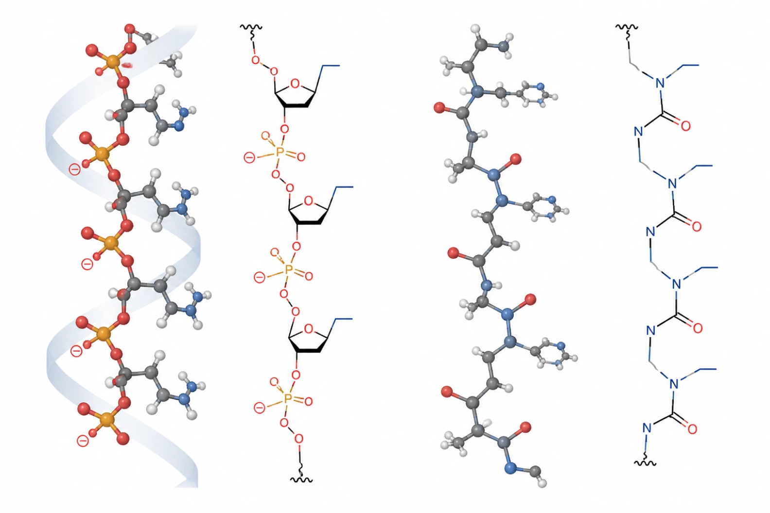
Biophysical Superiority of the PNA Backbone
The defining feature of a PNA molecule is its complete lack of electrostatic charge. In standard DNA-DNA or DNA-RNA duplexes, the negatively charged phosphate groups on opposing strands repel each other, requiring high concentrations of counter-ions (such as sodium or magnesium) to stabilize the double helix. The neutral pseudopeptide backbone of a PNA eliminates this electrostatic repulsion entirely.
Consequently, PNA-DNA and PNA-RNA duplexes exhibit significantly higher thermodynamic stability and melting temperatures ($T_m$) compared to their natural counterparts. A single base pair mismatch in a PNA-DNA duplex causes a more drastic drop in $T_m$ (often 10°C to 15°C) than an equivalent mismatch in a DNA-DNA duplex. This extreme binding affinity and mismatch discrimination dictate PNA's dominance in diagnostic applications requiring ultra-high specificity. Furthermore, because the backbone is structurally alien to natural biological systems, PNAs are highly resistant to degradation by both nucleases and proteases, granting them extended stability in crude biological samples, serum, and cellular lysates without the need for extensive sample purification.
PNA Synthesis Methods: SPPS Strategies
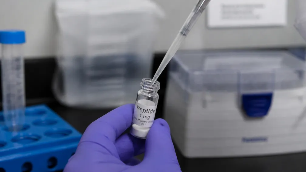
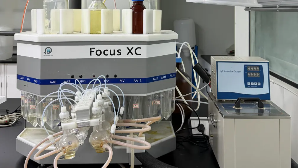
Given that the PNA backbone is composed of peptide bonds, the manufacturing process relies heavily on traditional custom peptide synthesis protocols rather than standard oligonucleotide phosphoramidite chemistry. Solid-Phase Peptide Synthesis (SPPS) is the undisputed industry standard for constructing PNA oligomers.
Fmoc-Mediated Synthesis
While early PNA synthesis relied on Boc (tert-butyloxycarbonyl) chemistry, Fmoc (9-fluorenylmethoxycarbonyl) chemistry has become the preferred industrial standard due to its milder deprotection conditions. Fmoc-SPPS eliminates the need for hazardous hydrofluoric acid (HF) during final cleavage, making it safer and more compatible with automated synthesizers and parallel manufacturing configurations. In Fmoc PNA synthesis, the nucleobase exocyclic amines are protected with acid-labile groups (e.g., Bhoc for adenine, cytosine, and guanine), which are cleaved simultaneously with the final release of the PNA from the resin using trifluoroacetic acid (TFA).
Coupling Reagents and Steric Hindrance
Coupling PNA monomers presents unique chemical challenges. The secondary amine of the N-(2-aminoethyl)glycine backbone introduces significant steric hindrance, reducing coupling efficiency compared to standard alpha-amino acids. To achieve quantitative yields, manufacturers must employ highly reactive coupling reagents, such as HATU (1-[Bis(dimethylamino)methylene]-1H-1,2,3-triazolo[4,5-b]pyridinium 3-oxid hexafluorophosphate) or HBTU, often combined with catalytic additives like HOAt or HOBt to suppress racemization and accelerate acyl transfer. Additionally, unreacted amines must be capped (typically using acetic anhydride) after each coupling cycle to prevent the formation of deletion sequences, which are notoriously difficult to separate during downstream purification.
Addressing Aqueous Solubility
A primary limitation of pure PNA oligomers is their high tendency to self-aggregate and poor aqueous solubility, particularly for sequences exceeding 15 bases or those with a high purine content. To circumvent this, chemical engineers routinely apply specific peptide modifications to the PNA construct. The most common strategy involves adding positively charged lysine residues (typically one to three) at the N- or C-terminus. Alternatively, incorporating hydrophilic polyethylene glycol (PEG) linkers, such as AEEA (aminoethoxyethoxyacetic acid), significantly enhances solubility and prevents non-specific binding in microfluidic diagnostic devices.
Purification and Analytical Quality Control
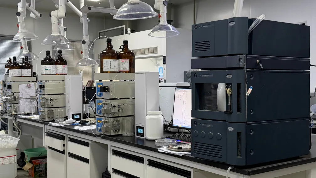
Purifying crude PNA mixtures requires robust analytical infrastructure. Reverse-Phase High-Performance Liquid Chromatography (RP-HPLC) is the standard method for isolating the target sequence from truncated failure sequences. Because PNAs lack the negative charge of DNA, traditional ion-exchange chromatography is ineffective. Optimizing the HPLC mobile phase is critical; PNAs typically require heating the column to 50°C–60°C to prevent secondary structure formation and self-aggregation during elution.
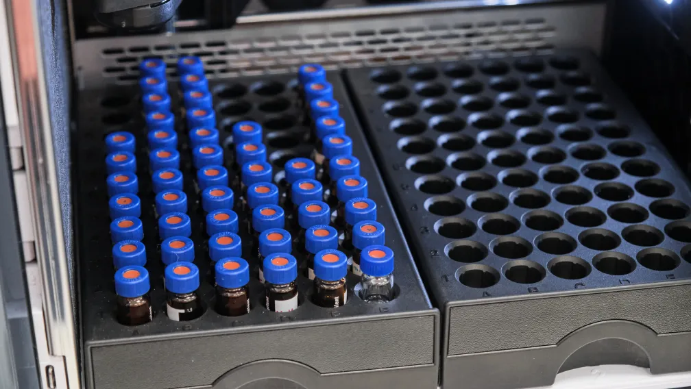
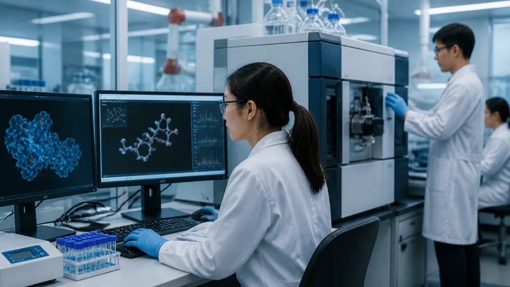
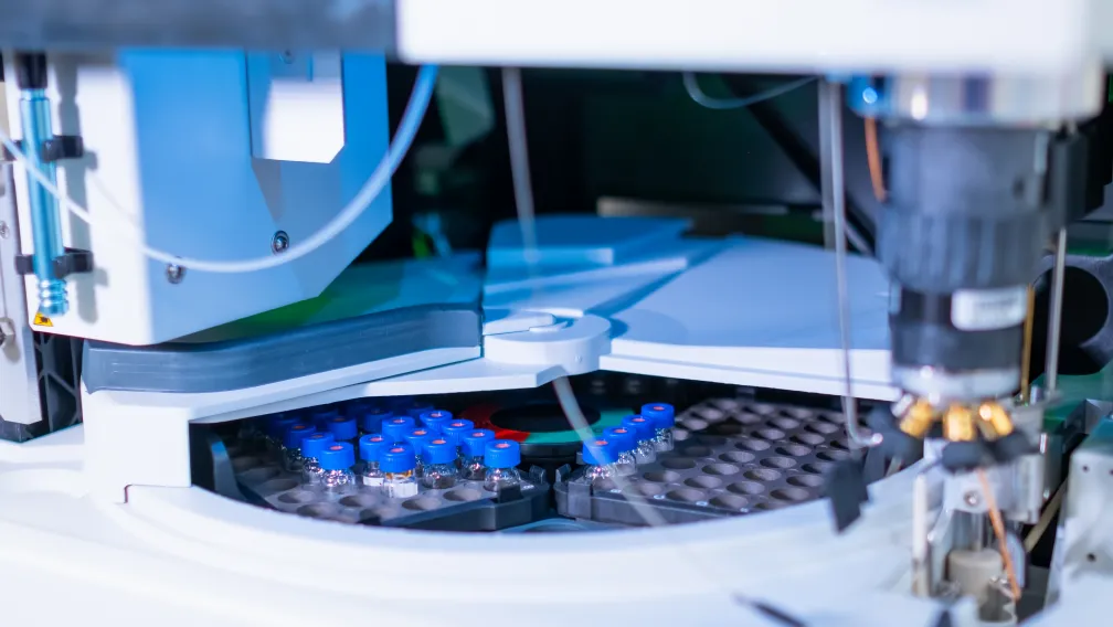
Sequence verification relies entirely on High-Resolution Mass Spectrometry (HRMS), specifically Liquid Chromatography-Mass Spectrometry (LC-MS) and Matrix-Assisted Laser Desorption/Ionization Time-of-Flight (MALDI-TOF). Ensuring sequence fidelity is paramount for catalog peptides and diagnostic probes, as a single truncation or incomplete deprotection step will obliterate the probe's binding kinetics and diagnostic utility.
Diagnostic Applications in Molecular Pathology
The superior hybridization kinetics of PNA probes have revolutionized in vitro molecular diagnostics, particularly in oncology, infectious disease identification, and cytogenetics.
PNA-PCR Clamping for Liquid Biopsies
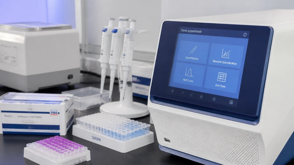
In clinical oncology, detecting rare mutant alleles (such as KRAS, EGFR, or BRAF mutations) in circulating tumor DNA (ctDNA) against a massive background of wild-type DNA is a monumental challenge. PNA-PCR clamping resolves this issue. A PNA probe is designed to perfectly match the wild-type DNA sequence. Due to its superior binding affinity, the PNA hybridizes tightly to the wild-type template. Because polymerases cannot recognize the PNA backbone, the PNA probe acts as an immovable physical clamp, entirely halting amplification of the wild-type sequence. Conversely, a single base mismatch at the mutation site drastically destabilizes the PNA-DNA duplex, allowing the mutant sequence to amplify freely. This method pushes the limits of detection down to 0.1% or lower, enabling highly sensitive liquid biopsy platforms.
Fluorescence In Situ Hybridization (PNA-FISH)
Traditional DNA-FISH assays are hampered by slow hybridization kinetics, requiring hours or overnight incubation. PNA-FISH probes, conjugated with fluorophores (such as FITC, Cy3, or Cy5), penetrate rigid bacterial cell walls and dense extracellular matrices much more efficiently than negatively charged DNA probes. The lack of electrostatic repulsion allows hybridization to occur in low-salt buffers within 30 to 90 minutes. This rapid turnaround time is critical in clinical microbiology, allowing for the direct, culture-independent identification of blood-borne pathogens (e.g., *Staphylococcus aureus*, *Candida albicans*) directly from positive blood cultures, dramatically accelerating targeted antibiotic administration.
Microarrays and Biosensors
In the development of next-generation biosensors, PNA probes are immobilized onto solid surfaces (such as gold nanoparticles, graphene oxide, or electrochemical electrodes). When target DNA or RNA binds to the immobilized PNA, it induces a measurable change in fluorescence, surface plasmon resonance, or electrical impedance. The uncharged nature of the PNA layer minimizes non-specific background noise from other charged biological molecules in crude serum or saliva samples, resulting in exceptionally high signal-to-noise ratios.
Scale-Up and CDMO Manufacturing Considerations
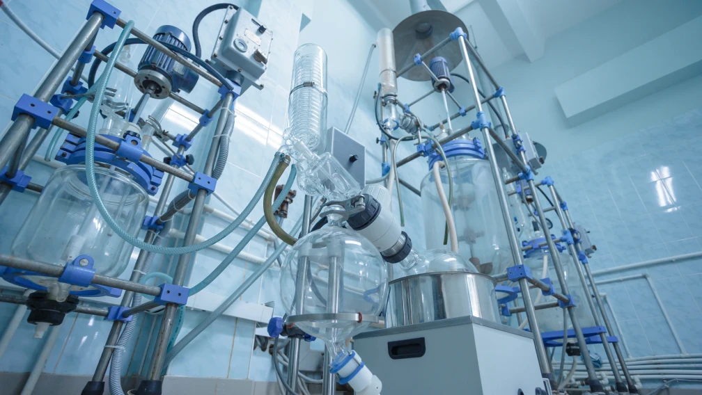
As diagnostic assays transition from laboratory research to commercial FDA-cleared or CE-IVD marked kits, the demand for high-purity PNA probes scales accordingly. Transitioning from nanomole synthesis to gram or kilogram quantities requires engaging a specialized peptide CDMO capable of large-scale peptide synthesis under strict ISO 13485 or GMP quality systems. Specialized manufacturing partners ensure that batch-to-batch variability is minimized, solvent usage is optimized to lower the Process Mass Intensity (PMI), and rigorous analytical testing guarantees that trace scavengers or guide to peptide modifications byproducts do not interfere with sensitive diagnostic enzymes.
Conclusion
Peptide Nucleic Acids have transcended their status as mere biochemical curiosities to become indispensable tools in the molecular diagnostic arsenal. By bridging the structural gap between proteins and nucleic acids, PNAs offer unparalleled binding affinity, mismatch discrimination, and chemical stability. For biotechnology developers, mastering the nuances of Fmoc-based PNA synthesis, solubility optimization, and analytical purification is the key to unlocking the next generation of ultra-sensitive genomic assays and rapid pathogen detection platforms.
-

Synpeptide
peptide-focused CRO/CDMO company
The SynPeptide Research Team brings together scientists specializing in peptide synthesis, purification, and analytical characterization. Drawing on hands-on laboratory experience across custom and catalog peptides, the team shares evidence-based insights for researchers, formulators, and product developers. All content is reviewed against current scientific literature and internal quality-control data, reflecting SynPeptide's commitment to accuracy, reproducibility, and the responsible communication of peptide science.